
 https://ibmm.umontpellier.fr/wp-content/uploads/2025/06/Morgan.jpg
226
400
Caroline Clavel
https://ibmm2.umontpellier.fr/wp-content/uploads/logo_ibmm_hd.jpg
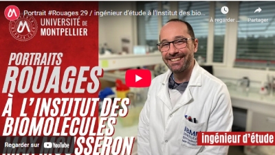
Caroline Clavel2025-06-19 16:40:542025-06-19 16:51:18Morgan Pellerano présente son métier sur le site de l’UM
https://ibmm.umontpellier.fr/wp-content/uploads/2025/06/Morgan.jpg
226
400
Caroline Clavel
https://ibmm2.umontpellier.fr/wp-content/uploads/logo_ibmm_hd.jpg
Caroline Clavel2025-06-19 16:40:542025-06-19 16:51:18Morgan Pellerano présente son métier sur le site de l’UMActualités
https://ibmm.umontpellier.fr/wp-content/uploads/2025/06/Morgan.jpg
226
400
Caroline Clavel
https://ibmm2.umontpellier.fr/wp-content/uploads/logo_ibmm_hd.jpg
Caroline Clavel2025-06-19 16:40:542025-06-19 16:51:18Morgan Pellerano présente son métier sur le site de l’UM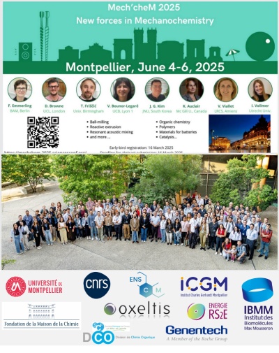 https://ibmm.umontpellier.fr/wp-content/uploads/2025/06/Image2.jpg
505
400
Caroline Clavel
https://ibmm2.umontpellier.fr/wp-content/uploads/logo_ibmm_hd.jpg
Caroline Clavel2025-06-13 11:49:342025-06-13 11:52:56Grand succès pour le congrès Mech’cheM !
https://ibmm.umontpellier.fr/wp-content/uploads/2025/06/Image2.jpg
505
400
Caroline Clavel
https://ibmm2.umontpellier.fr/wp-content/uploads/logo_ibmm_hd.jpg
Caroline Clavel2025-06-13 11:49:342025-06-13 11:52:56Grand succès pour le congrès Mech’cheM !Évènements
Stages / emplois
16 équipes de recherche

Polymères pour la Santé et Biomatériaux

Chimie Verte et Technologies Innovantes

Oncopharmacochimie & Pharmacotoxicologie Cutanée

Oncothérapie et oncopharmacologie

Dynamique des Systèmes Biomoléculaires Complexes

Glyco et Nanovecteurs pour le ciblage Thérapeutique

Sciences analytiques des biomolécules

Synthèse stéréosélective & acides aminés modifiés

Acides aminés, hétérocycles, peptides & protéines

Pharmacologie Cellulaire

ChemBioNAC
Pharmacochimie, transmission synaptique & neuroprotection

Supramolecular MAchines and ARchitecture Team

Synthèse de Lipides Bioactifs

Nucléosides & Effecteurs Phosphorylés

Offre de thèse : PHOTOCLICK: Development of biorthogonal photo-crosslinking reactions for 3D printing micro-structured biomaterials
Contract: 3 years starting from October 2025 Application deadline: 04/07/2025 Location: Institute of Biomolecules Max Mousseron (IBMM), Montpellier, France and Institute for Molecular Systems Engineering and Advanced Materials (IMSEAM), Heidelberg, Germany The project This thesis project is located at the interface of click chemistry, photochemistry, materials, 3D printing and biology. The goal is to […]
Offre de thèse : Advanced 4D biomaterials for mucosa and sub-mucosa treatment in patients affected by intestinal diseases (Daedalus)
ADVANCED 4D BIOMATERIALS FOR MUCOSA AND SUB-MUCOSA TREATMENT IN PATIENTS AFFECTED BY INTESTINAL DISEASES (Daedalus) financed by the EU program HORIZON EUROPE 4D biomaterials enable new surgical treatments by autonomously acting in response to environmental stimuli, thus overcoming the limitations of standard medical strategies. DAEDALUS is a European project that gathers 13 partners and […]